Anatomy Of The Liver
The liver carries out highly specialized processes that are essential for health and life, including detoxification processes, protein synthesis, and so on.
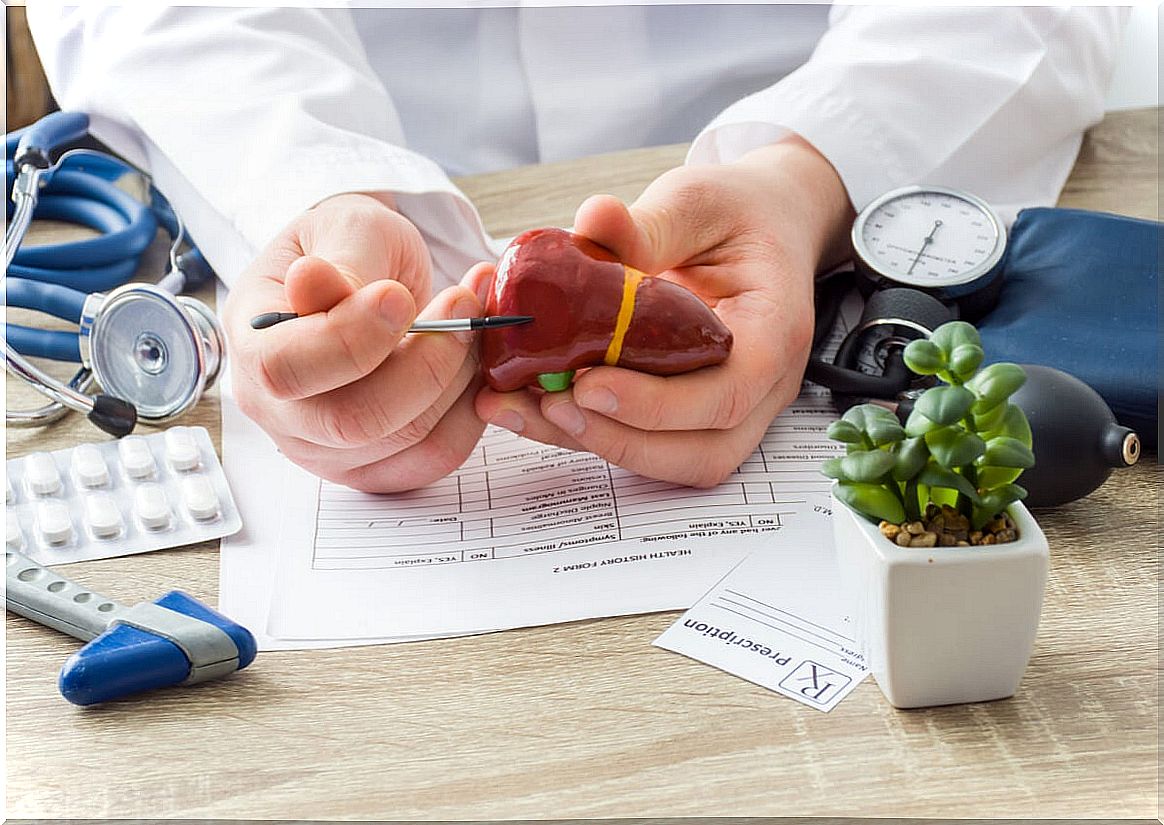
The liver is a gland attached to the digestive system that is present in all vertebrates. Many call it the “body’s laboratory” because it performs very complex processes to purify the blood.
It is one of the organs that intervenes in digestion, especially in that of fats through bile. In addition, it has many other functions, including the synthesis of plasma proteins, storage of vitamins and glycogen and detoxifying function.
Do you want to know more about this organ? Keep reading everything that we are going to tell you about!
The curious history of the word “liver”
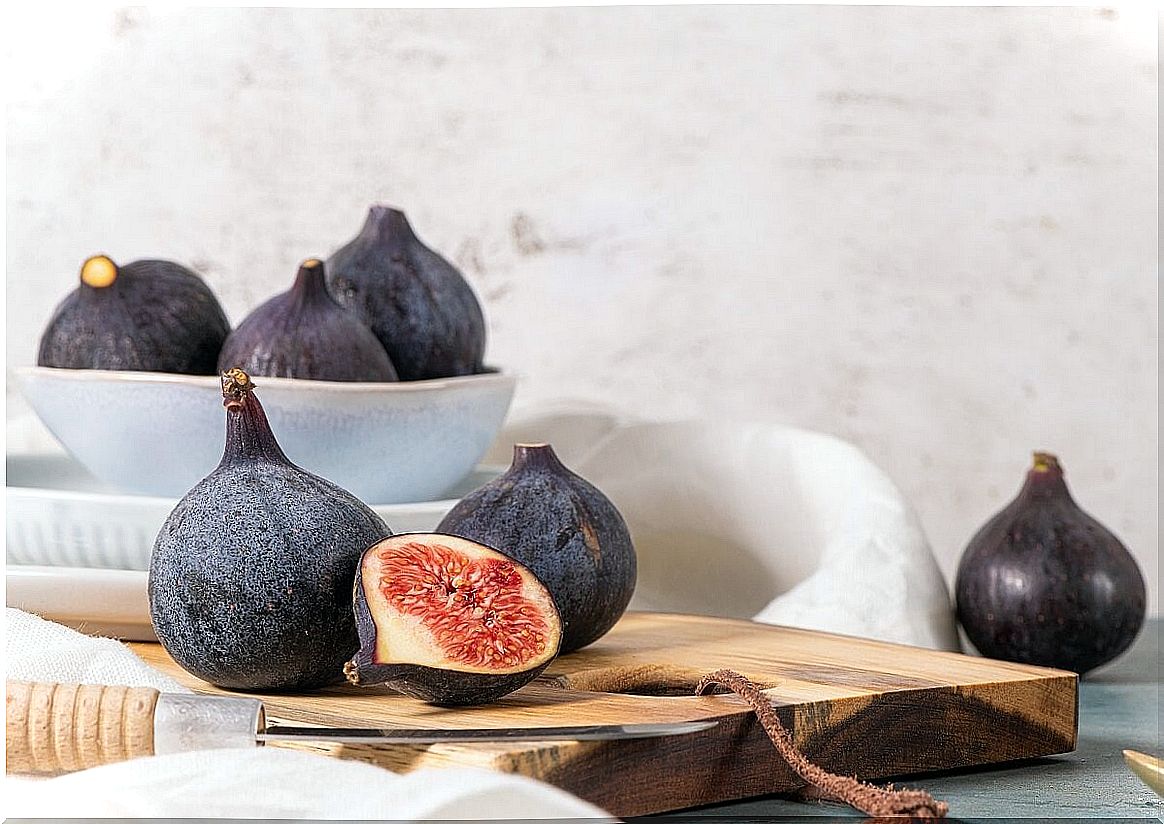
As the University of Salamanca Medical Dictionary indicates, the term “liver” has a curious origin. It comes from the Latin term ficatum, which in turn is derived from ficus , that is, ‘fig’. This curious association of words came about because the Romans fattened geese with figs. This caused the liver to swell in these animals, which some consider to be a gastronomic delight.
But not only its name is interesting. The scientific literature reveals data as striking as:
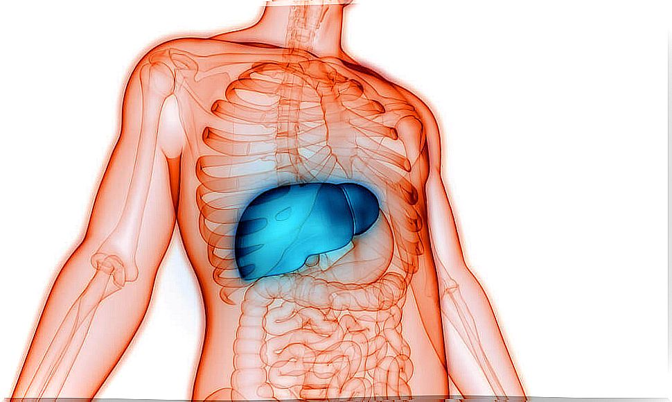
- The liver is located in the upper right part of the abdominal cavity.
- It has the appearance of a dark reddish-brown cone, because it is filled with blood.
- It is the largest organ in the body. It can weigh about 1400 grams in an adult woman and 1800 in an adult man. This means that it represents 2% of body weight.
The location and appearance of the liver
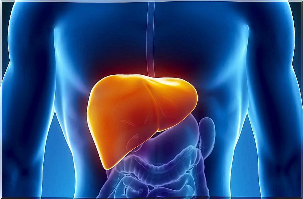
The liver is located below the diaphragm, above the duodenum (small intestine), and in front of the stomach. It occupies the right upper quadrant and a portion of the epigastrium. It is very close to the heart, but is separated from this organ by the diaphragm. It has two faces and an edge.
- Diaphragmatic face. It is made up of the “anterior superior” face, or the one located at the top and from the front; and an “extraperitoneal” zone, in the posterior part. The latter is outside the peritoneum, that is, the membrane that covers most of the viscera. This face is divided into right and left, thanks to the “falciform ligament”.
- Visceral face. It comprises a lower face and an area lined with peritoneum, in the posterior part. On this face there are three grooves arranged in the shape of an “H”. These are: gallbladder fossa, round ligament fissure, and portahepaticus. The grooves, in turn, make it possible to isolate four lobes: right, square, left, and caudate.
The diaphragmatic face and the visceral face join at the lower border. At the base of the liver is the gallbladder and the hepatic hilum. The latter is the entry zone of the portal vein and the hepatic artery. It is also the exit point of the hepatic duct.
Couinaud’s classification
Claude Couinaud made a classification according to which the liver is divided into eight parts. Each of them is independent. These segments are numbered according to clockwise movement and are as follows:
- In the right liver lobe : there are segments VII and VIII at the top and VI and V at the bottom.
- The left liver lobe contains segment II (subdiaphragmatic), IV (very medial), and III (below II).
- In the caudate lobe is the segment I, but is frontally visible.
Each segment has a branch of the hepatic artery. In turn, a branch of the hepatic vein emerges from each and also a branch of the portal vein arrives.
Ligaments of the liver
The liver is almost completely lined by the visceral peritoneum. At the same time it has several connections with the parietal peritoneum through various fibrous tracts, ligaments:
- Falciform.
- Coronary.
- Round liver.
- Gastrohepatic.
- Venous duct.
- Hepatoduodenal.
All ligaments together constitute a means of fixation. This means that they support the liver, that is, they keep it in position. They also fulfill the function of supporting the organ on the adjoining structures.
The vessels and nerves of the liver
The liver receives arterial blood through the hepatic artery. This blood goes to the parenchyma, that is, the essential and functional tissue of the organ. On the other hand, the venous blood, coming from the abdominal viscera, passes through the portal vein.
Between 70% and 75% of the non-oxygenated blood that circulates through the liver is carried by the portal. Its tributaries are: the right gastric vein, the left, the posterior superior pancreatoduodenal, the prepyloric, the paraumbilical veins and those coming from the bile ducts.
A redoubt of the venous blood is transported by the retroperitoneal veins. Also, during pregnancy, the blood of the fetus runs through the umbilical vein, from the placenta.
Arterial and venous blood mix in capillaries called “hepatic sinusoids. ” Then the mixed blood leaves the organ through the hepatic veins, also known as “suprahepatic”, which flow into the inferior cava.
